


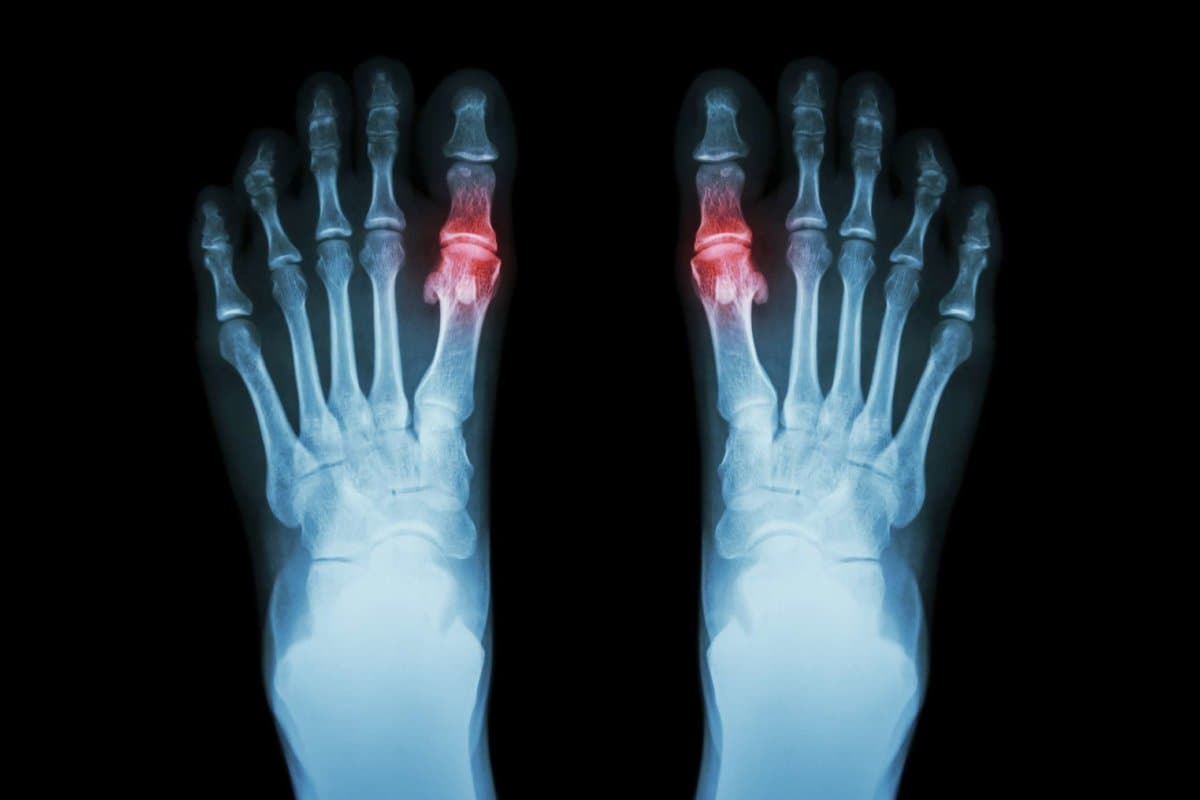
Gout can be diagnosed clinically during an acute attack. In addition, imaging provides valuable information about the condition of joints that are not severely affected and about the damage that has already occurred. Recommendations for the use of imaging in gout and other crystalline arthropathies have now been summarized in a guide presented at the EULAR 2023 conference.
stockdevil/GettyImages
Crystal arthropathies (crystalline-induced arthropathies – CiA) represent a diverse group of diseases in which deposition of crystalline metabolites occurs in the joint cavity and subsequent arthritis. Uric acid or sodium urate (gout), calcium pyrophosphate crystals (chondral calcification), hydroxyapatite crystals (hydroxyapatite deposition disease) and homogenous acid (synchronous arthropathy) can crystallize in the joint cavities. Imaging can play an important role in the diagnosis of crystalline arthropathies, but it is by no means as trivial in this indication as Priv.-Doz. Dr. Peter Mandel of the Vienna University Clinic for Internal Medicine III. Diseases present clinically with acute episodes of arthritis, followed by asymptomatic episodes.
In its guidelines for the use of imaging in the diagnosis and follow-up of crystalline arthropathies, the EULAR Task Force has created guidelines with evidence-based recommendations. The task force consisted of 25 people from 11 countries with different professional and academic backgrounds. Four systematic literature searches of MEDLINE, EMBASE, and CENTRAL were designed to support the task force’s decisions on 14 questions, considering the indications for gout, chondrocalcinosis, and hydroxyapatite deposition disease.

Sylvia Plath writes for Social Post News, covering news, politics, business, technology, sport, entertainment, and lifestyle. She focuses on clear, reliable reporting and useful information, helping readers stay informed about current events, emerging trends, and stories that matter.


More Stories
Bumblebees Show Problem-Solving Skills Once Thought Limited to Larger-Brained Animals
Artemis 2’s Orion Heat Shield Faces Critical Test as Astronauts Prepare for Fiery Earth Reentry
Coral Seeding: Artificial Insemination Makes Coral More Heat Tolerant